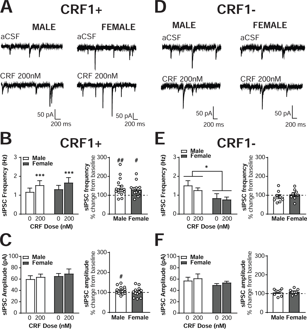
Figure 2. CRF increases sIPSC frequency in male and female CRF1+, but not CRF1-, CeA neurons.
(A) Representative voltage-clamp recordings from male (left) and female (right) CRF1+ CeA neurons during focal application of aCSF (top) or CRF (200nM; bottom). (B) Quantification of sIPSC frequency in male and female CRF1+ CeA neurons following aCSF or CRF (200nM) focal application expressed in Hz (left) and percent of control (right). (C) Quantification of sIPSC amplitude in male and female CRF1+ CeA neurons following aCSF or CRF (200nM) focal application expressed in picoamps (left) and percent of control (right). (D) Representative voltage-clamp recordings from male (left) and female (right) CRF1- CeA neurons during focal application of aCSF (top) or CRF (200nM; bottom). (E) Quantification of sIPSC frequency in male and female CRF1- CeA neurons following aCSF or CRF (200nM) focal application expressed in Hz (left) and percent of control (right). (F) Quantification of sIPSC amplitude in male and female CRF1- CeA neurons following aCSF or CRF (200nM) focal application expressed in picoamps (left) and percent of control (right). * = p < 0.05 by two-way RM ANOVA, *** = p < 0.001 by two-way RM ANOVA, # = p < 0.05 by one-sample t-test, ## = p < 0.01 by one-sample t-test.

