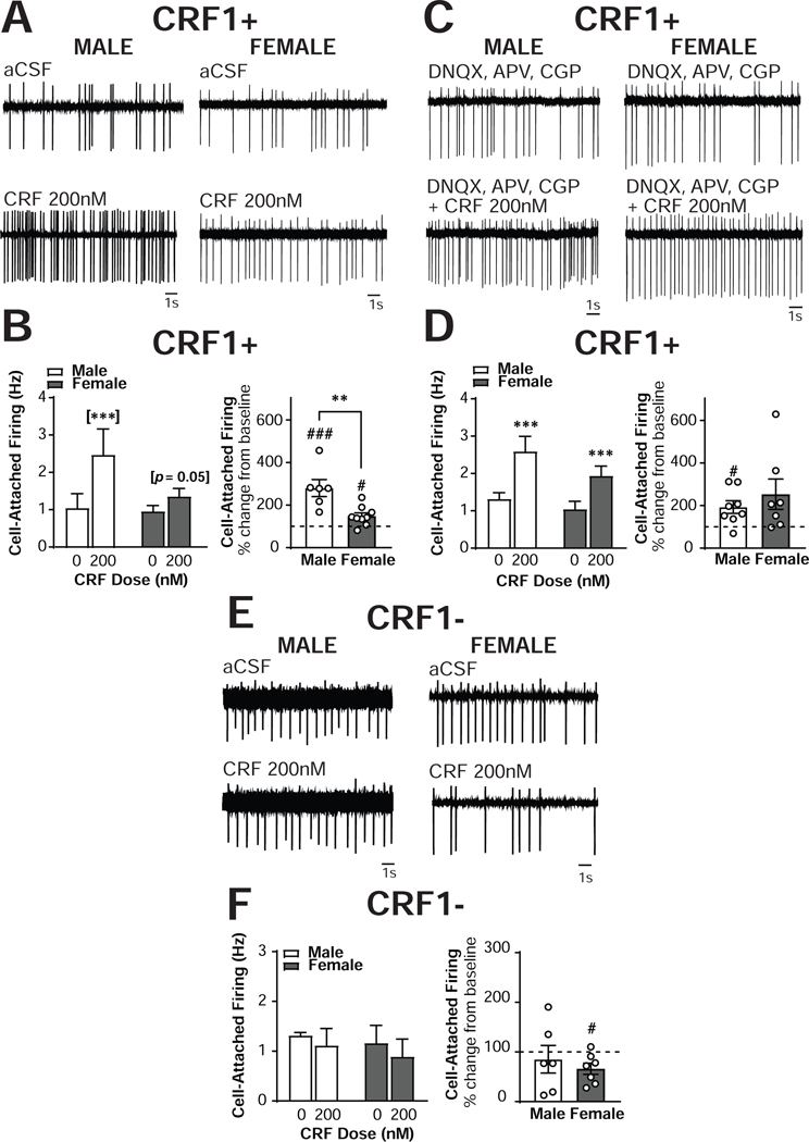
Figure 6. Sex differences in CRF regulation of excitability in CRF1+, but not CRF1-, CeA neurons.
(A) Representative cell-attached recording of male (left) and female (right) CRF1+ CeA neurons during focal application of aCSF (top) and CRF (200nm; bottom). (B) Quantification of cell-attached firing frequency in male and female CRF1+ CeA neurons following focal application of aCSF and CRF (200nM) expressed in Hz (left) and percent of control (right). (C) Representative cell-attached recording of male (left) and female (right) CRF1+ CeA neurons during bath superfusion of DNQX, APV and CGP with focal application of aCSF (top) and CRF (200nm; bottom). (D) Quantification of cell-attached firing frequency in male and female CRF1+ CeA neurons following focal application of aCSF and CRF (200nM) in the presence of DNQX, APV and CGP expressed in Hz (left) and percent of control (right). (E) Representative cell-attached recording of male (left) and female (right) CRF11 CeA neurons during focal application of aCSF (top) and CRF (200nm; bottom). (F) Quantification of cell-attached firing frequency in male and female CRF1- CeA neurons following focal application of aCSF and CRF (200nM) expressed in Hz (left) and percent of control (right). ** = p < 0.01 by two-sample t-test, # = p < 0.05 by one-sample t-test, ## = p < 0.01 by one-sample t-test, [***] = p < 0.001 by Sidak’s multiple comparison’s test.

