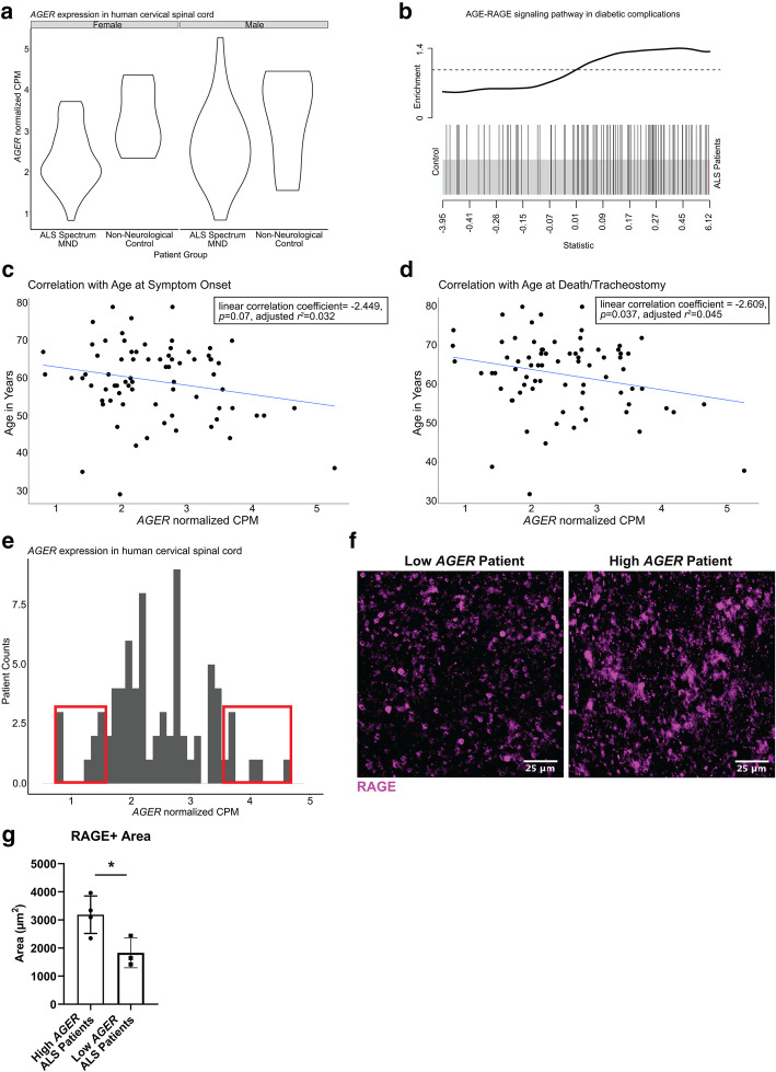
Fig. 1.
Analysis of human ALS patient RNA-seq data implicates the RAGE pathway in modulating disease. a Normalized AGER counts per million mapped reads across patient groups separated by sex (no significant sex-dependent differences). b Barcode plot illustrating gene set enrichment of the “AGE-RAGE signaling pathway in diabetic complications” between ALS patients and non-neurological controls. The snake illustrates the enrichment and upregulation of the gene set in ALS patients. c Correlation of AGER expression (x-axis) and age at onset in years (y-axis). Regression line shown. Coefficient = − 2.449, p = 0.07, adjusted r2 = 0.032. d Correlation of AGER expression (x-axis) and age at death/tracheostomy in years (y-axis). Regression line shown. Coefficient = − 2.609, p = 0.037, adjusted r2 = 0.045. e Histogram of normalized AGER expression in ALS patients. Red boxes notate the highest 10% and the lowest 10% of AGER-expressing patients. f Representative image of regions of interest for RAGE staining in the ventral horn of a high and low AGER patient cervical spinal cord. Scale bar: 25 μm. g Quantification of RAGE+ area. In a: from left to right N = 38, 5, 38, 6. In b: N = 76 ALS patients and N = 11 non-neurological control patients. In c–e: N = 76 ALS patients. In f, g: N = 4 high AGER and 3 low AGER patients. Independent two sample two-sided t test. *p = 0.0349

