Fig. 10.
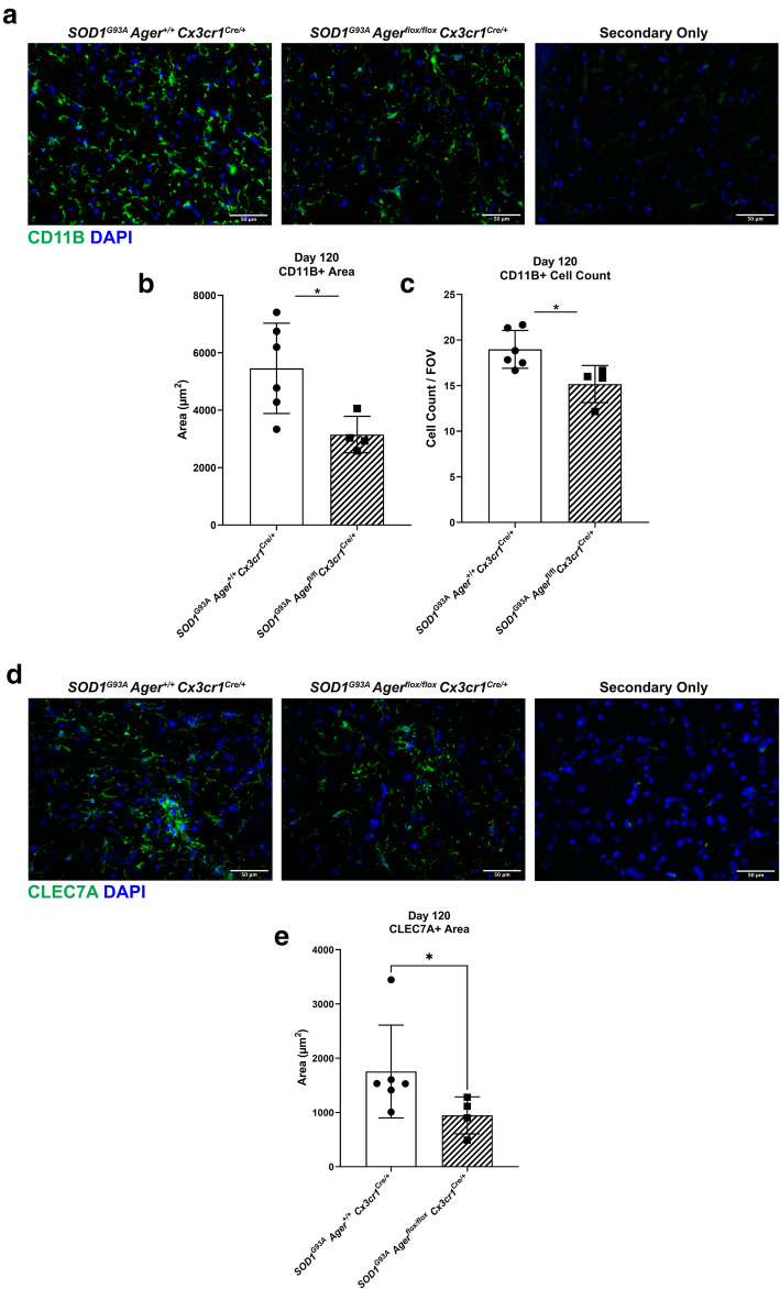
Microglia Ager deletion in male SOD1G93A mice reduces microgliosis at 120 days of age. a Representative images of CD11B staining in the lumbar spinal cord ventral horn of the indicated mouse groups. b Quantification of CD11B+ area. c. Quantification of CD11B+ DAPI+ cell number. d Representative images of CLEC7A staining in the lumbar spinal cord ventral horn of the indicated mouse groups. e Quantification of CLEC7A+ area. Mean ± SD. N = 4 SOD1G93A Agerfl/fl Cx3cr1Cre/+ mice, N = 6 SOD1G93A Ager+/+ Cx3cr1Cre/+ mice. In b, c, independent two sample two-sided t test. In e, Mann-Whitney U test. In b, * p = 0.0253. In c, *p = 0.0212. In e, *p = 0.0381

