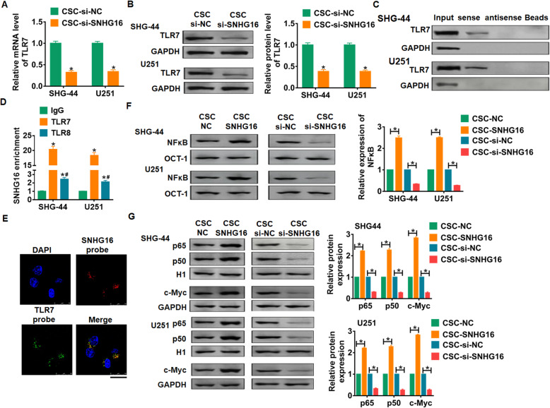
Fig. 4.
SNHG16 activated TLR7- NFκB-c-Myc signaling pathway. SHG44 and U251 cells were incubated with exosomes isolated from CSCs transfected si-SNHG16/si-NC. A qRT-PCR analyzed the expression of TLR7 in SHG44 and U251 cells. n = 6, *p < 0.05. B Western blot for TLR7 expression in SHG44 and U251 cells. n = 4, *p < 0.05. C Western-blotting validation of TLR7 in SNHG16 pulldown protein extractions. D RNA-immunoprecipitation experiments were performed using TLR7, TLR8 and IgG antibodies to immunoprecipitate SNHG16 in SHG44 and U251 cells. E FISH assay was used to detected the subcellular location of SNHG16 and TLR7 in U251 cells, the nucleus was stained using DAPI. Scale bar = 25 μm. F NFκB activities was examined by EMSA. n = 4, *p < 0.05. G Western blotting showing nuclear p65, p50, Histone H1 and c-Myc levels in SHG44 and U251 cells. n = 4, *p < 0.05

