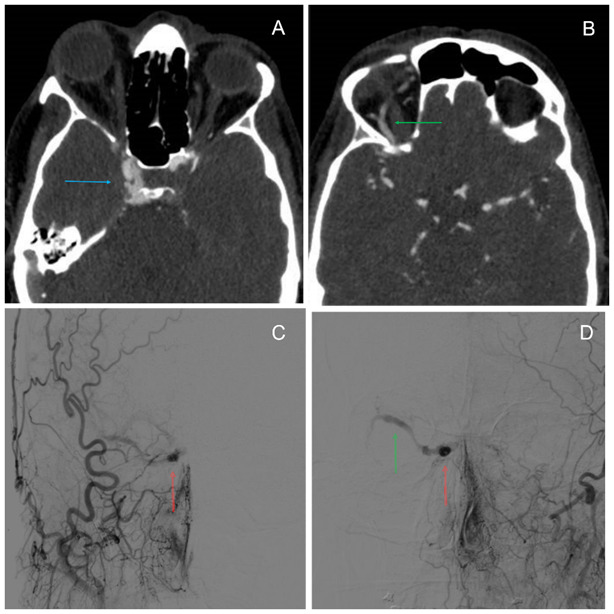
Fig. 2.

A. Angio-CT scan: in the artery phase, an image of early and abnormal filling of the right cavernous sinus (CS) was seen (blue arrow). B. Enlarged superior ophthalmic vein (SOV) characteristic of Carotid Cavernous Fistula (CCF) (green arrow). C and D. Brain arteriography revealed the abnormal filling of the right CS (red arrow) and enlarged SOV (green arrow) from right and left internal carotid artery respectively. Carotid Cavernous Fistula Type B Barrow Classification
