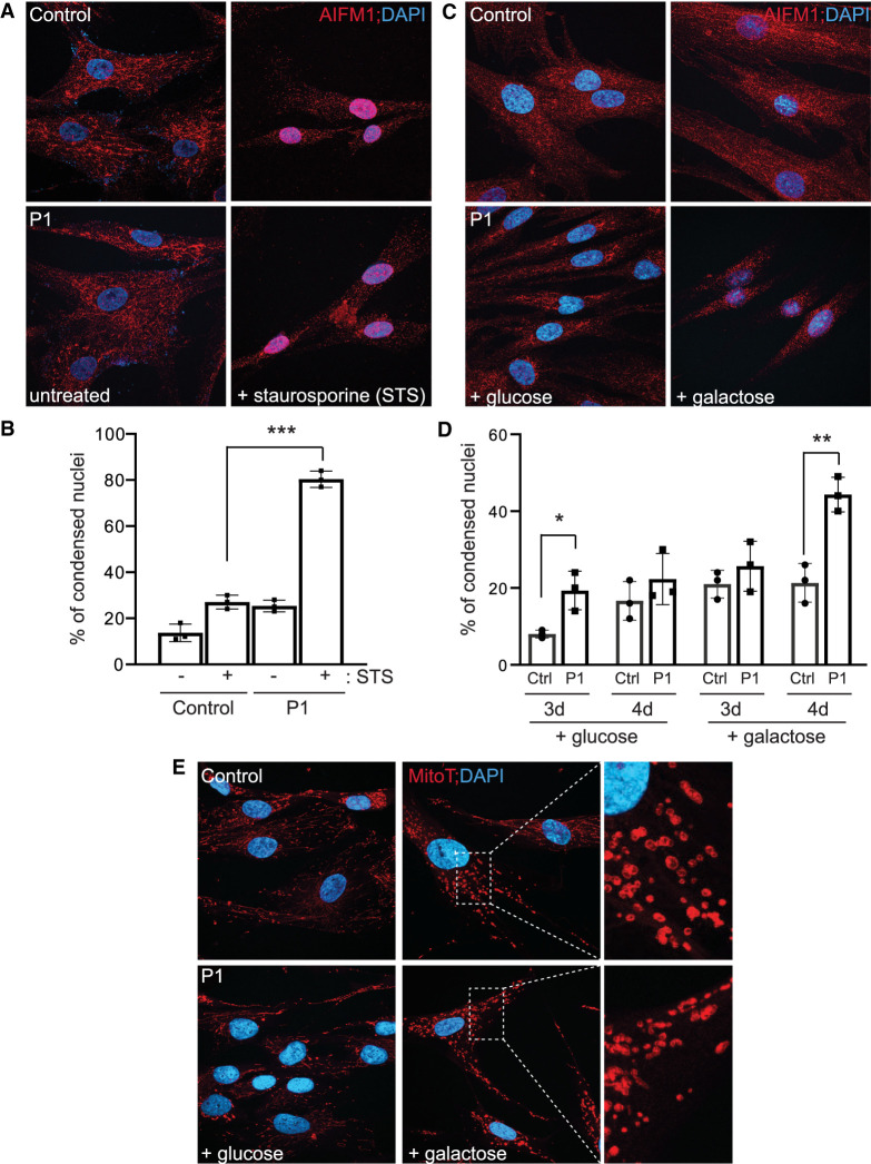
Figure 3.
Increased sensitivity of patient fibroblasts to the apoptotic inducer staurosporine. (A) Immunostaining of control and P1 fibroblasts using an antibody to AIFM1 before and after treatment with staurosporine. DAPI is used as a nuclear marker. (B) Quantification of the number of condensed nuclei in untreated and staurosporine-treated control and P1 fibroblasts. *** P < 0.001. (C) Representative images of AIFM1 immunostaining in wild-type (WT) and patient fibroblasts cultured in glucose- or galactose-containing media for 96 h. (D) Quantification of the percentage of condensed nuclei under these growth conditions for both 72 and 96 h. (E) Representative images of MitoTracker CMX ROS Red staining in cells incubated with glucose or galactose for 96 h. Enlarged inset is provided to highlight the morphology of the fragmented mitochondria.

