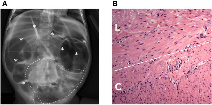
Figure 1.

(A) Abdominal radiograph shows severe diffuse gaseous distension of bowel throughout the abdomen (asterisks). Scattered bubbly lucencies (within dashed line) may represent stool contents. Enteric tube and peripherally inserted central catheter (PICC) tip are also visible. (B) Hematoxylin and eosin (H&E) staining of the patient's small bowel (ileostomy) specimen reveals no abnormalities in smooth muscle band thickness or organization. (L) Longitudinal layer, (C) circular layer.
