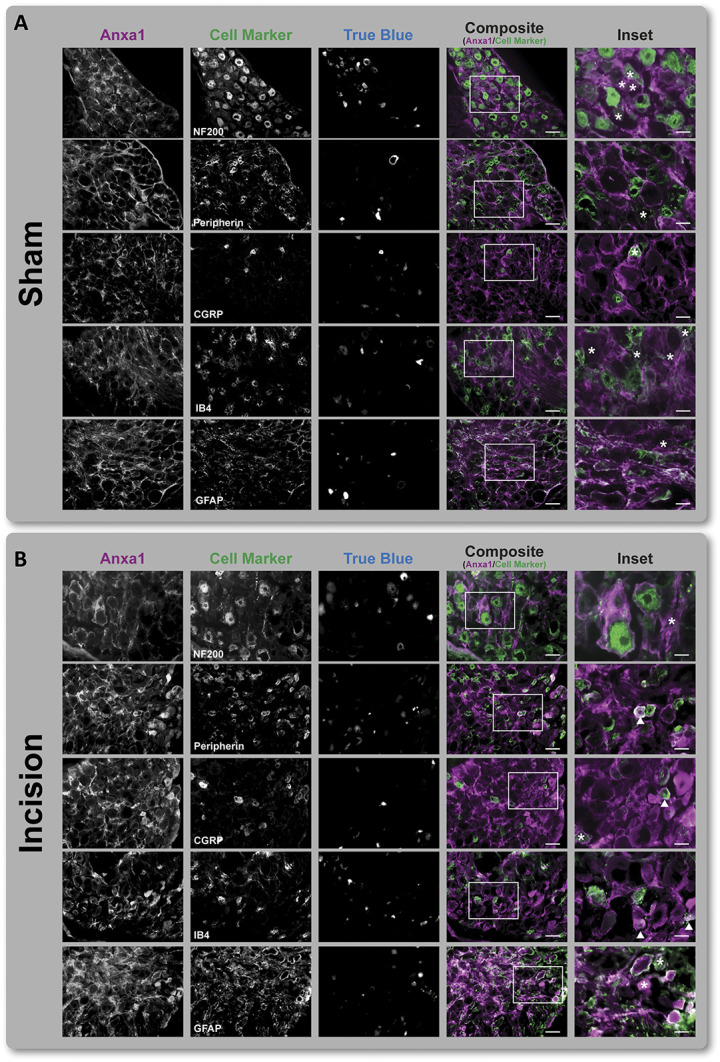
Figure 6.

Localization of Anxa1 in L4 ipsilateral DRG. (A) Under Sham conditions, Anxa1 is colocalized with GFAP-positive cells (ie, satellite glia cells) but not with neuronal subpopulations (labeled by indicated marker proteins) or retrogradely labeled neurons (True Blue positive) in ipsilateral L4 DRG. (B) By contrast, plantar incision (postoperative day 1) induced neuronal expression of Anxa1 in Peripherin-, CGRP- and IB4-positive neurons (indicated by arrows, Inset). Retrogradely labeled neurons are marked with an asterisk (Inset). Scale bar = 50 µm (composite), 20 µm (Inset). DRG, dorsal root ganglia.
