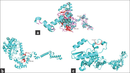
Figure 4.

Modeled structures of normal and mutant XPC proteins. (a) Structure of wild type XPC protein, the arrows show the position of frameshift start in diseased individuals; (b) structure of p.P462Tfs*32 mutant protein, the miss-sequence residues (461-491) are shown in red color; (c) structure of p.P645Lfs*5 mutant protein, the miss-sequence residues (645-648) are shown in red
