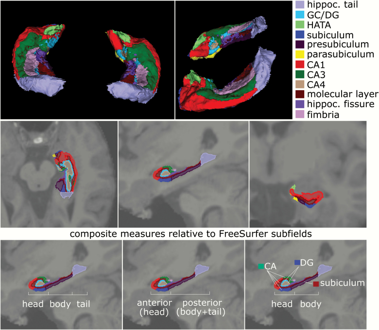
Fig. 1.
Description of hippocampal subfields derived from original FreeSurfer 6.0 segmentation. Top row shows 3D model of the different segmented hippocampal subfields with top and lateral views on the segmented hippocampus (color legend of individual subfield segments on right); middle row shows transverse, sagittal, and coronal sections of the hippocampus with subfields superimposed on T1-weighted MRI scan; the bottom row shows the relation of these original FreeSurfer 6.0 hippocampal subfields to functional models separating the hippocampus into either (from left to right) head, body, and tail components, or an anterior vs posterior part, or finally a model grouping cornu ammonis (CA), dentate gyrus (DG), and subiculum subfields within the head-body separation.

