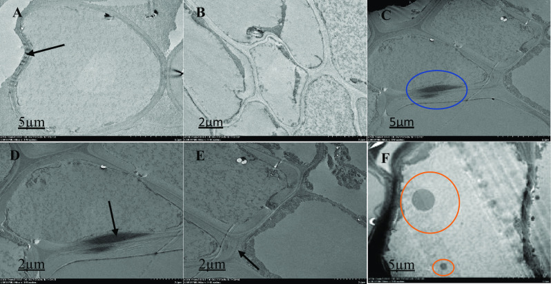
Fig. 2.

Ultrastructural organization of M. malabathricum roots, TEM images showing the cell wall, nuclear membrane, nucleus and vacuole. Tissue samples were obtained after 90 days of exposure to 50 ppm As + bacteria in soil. Thick arrow shows cell wall (A, D and E), blue circle showing unidentified image (C), red circle shows electron dense material (F), higher magnification observed cell structure: extracellular dark deposits are observed in several cells in contact with the outer face of the cell walls, some two-cell structures showing different asymmetric location of the dividing cell wall and some specifics of the organization of the cell (B)
