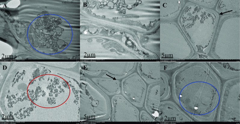
Fig. 4.

Ultrastructural organization of M. malabathricum roots, TEM images showing the cell wall, nuclear membrane, nucleus and vacuole. Tissue samples were obtained after 90 days of exposure to 50 ppm As in soil. Thick arrow shows cell wall (C and E), blue circle shows unidentified structure (A, B and F), red circle shows electron dense material (D). Higher magnification of the specifics of the cell organization; some two-cell structures showing the dividing cell wall's distinct asymmetrical location and certain cell organization features. Chloroplasts with denser-structured internal membranes showed a rounded shape and smaller scale
