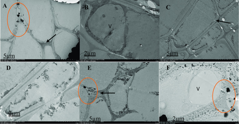
Fig. 5.

Ultrastructural malabathricum root organization, TEM photos illustrating the cell wall, nuclear membrane, nucleus, and vacuole. After 90 days of exposure to 70 ppm of arsenic in the soil, tissue samples were taken. Thick arrow shows cell wall (A, C and E), red circle shows thick electron content (A, D and F), V = vacuole (B). Higher magnification of the specifics of the cell organization; extracellular dark deposits (red circle) are found in many cells in contact with the outer face of the cell walls; cells show large vacuoles (V). On the cell walls, extracellular deposits of various sizes and types (rounded and ellipsoid) are found
