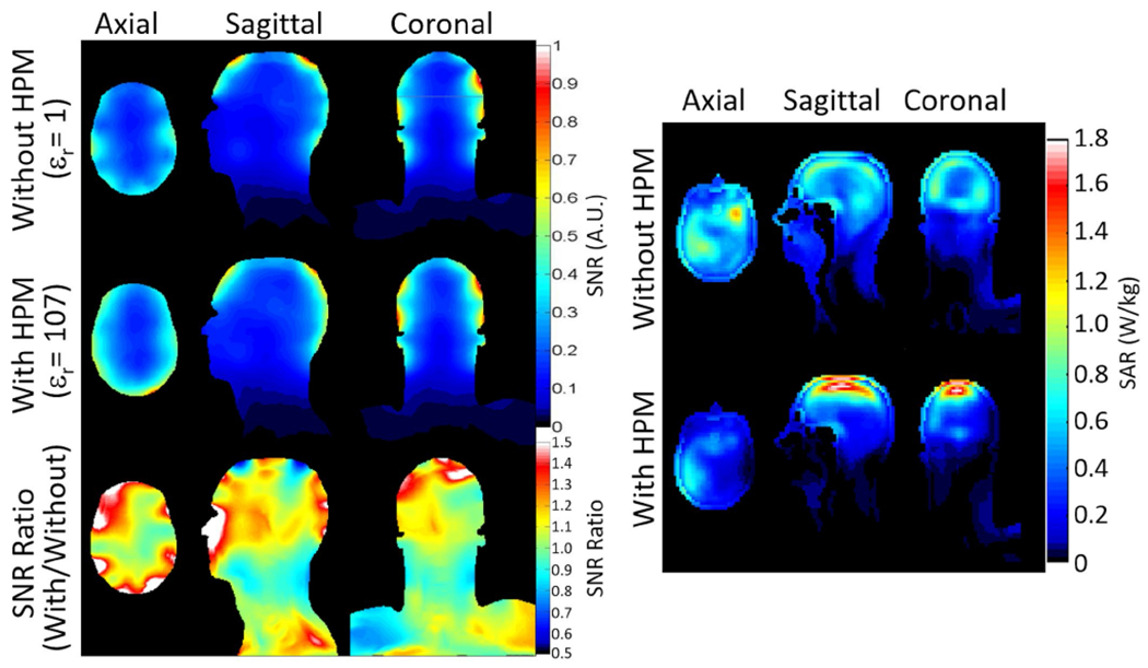
FIGURE 4.

Left: Simulated SNR in a 30-channel array as distributed through the head on three orthogonal planes for 30-channel receive array without HPM and with coils moved closer to the head (top row), with HPM (middle row), and the ratio of SNR with HPM to that without (bottom row). Right: Distribution of 10-g average SAR for 1 μT|| at center of brain for dipole array without (top) and with (bottom) HPM on axial plane through maximum 10-g SAR for case without HPM and on sagittal and coronal planes through maximum 10-g SAR for case with HPM
