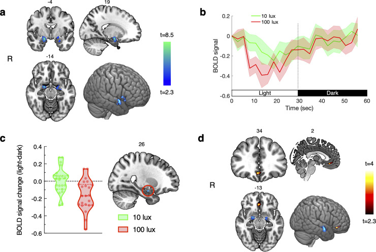
Fig 1. Light, compared with dark, decreased activation in the amygdala and increased functional connectivity between the amygdala and ventro-medial prefrontal cortex.
(a) Voxels with significantly decreased activity in the amygdala during light relative to dark (p<0.05, small volume correction using bilateral amygdala mask); (b) average time-course of the baseline-corrected BOLD signal across individuals (shaded areas represent SEM). Time-courses were obtained by averaging the normalized BOLD signal for the 8 cycles each of light and dark periods from significant voxels within the amygdala; (c) BOLD signal % change in the cluster (4 voxels) showing greater deactivation during 100 lux compared with 10 lux (p<0.05, cluster corrected within the bilateral amygdala mask). The peak voxel was located at MNI: 25, -7, 19. Individual responses are represented by circles, and dashed and dotted lines represent the median and upper/lower quartiles, respectively; and (d) voxels (53 voxels) within the vmPFC mask showing a significant interaction effect for light vs. dark for functional connectivity with the amygdala (p<0.05, cluster corrected within the vmPFC mask). The peak voxel was located at MNI: 2, 35, -14. Note: Slice labels in panels a, c, and d denote MNI slice numbers. Activation maps are shown on an MNI template and visualized using MRICroGL software using radiological orientation.

