
Figure 8.
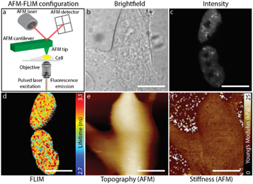
Correlative AFM and FLIM imaging of neuron like SHSY-5Y cells expressing the intranuclear protein FUS labelled with YFP. Aberrations in the phase transitioning behaviour of FUS is associated with neurodegenerative disease such as amyotrophic lateral sclerosis, ALS. Panel (a) shows a schematic of the AFM-FLIM setup on an inverted microscope. (b) Bright field image of the cell outlines. (c) Fluorescence image of FUS-YFP, demonstrating the localisation of the protein to the nucleus. (d) FLIM image of FUS-YFP which informs on the phase state of the protein. (e) and (f) Show images obtained by AFM informing on cell topology and cell stiffness, respectively. Together, the mechanical and lifetime data inform on relationships between cellular phenotypes and phase transitioning behaviour of FUS. Scale bar: 10 μm.