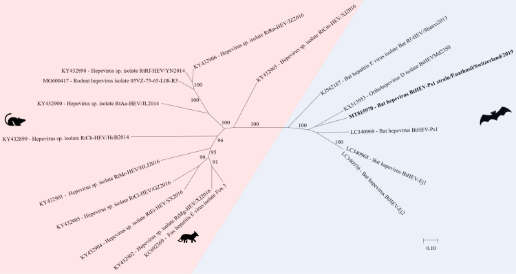
Fig 11. Phylogenetic analysis of full and partial genome of hepeviruses.
The sequence obtained in this study (GenBank acc. numbers MT815970) is shown in bold. Sequences of non-bat associated viruses are marked with a red background and those of bat associated viruses with a blue background. The pictograms on the right side represent the species in which the virus was detected. Sequences were aligned using Muscle. For phylogenetic analysis, the Maximum likelihood tree with 1’000 bootstraps was used. Only values ≥ 70% are displayed.

