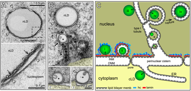
Fig 5. Membrane-enclosed nLDs.
(A) TEM of tubule-associated nLD with extra membranes (white arrowheads) in a D2 nucleus; see also Fig 4C and 4D. The high magnification inset (bottom) shows that the nLD surface (black arrows) is partially surrounded by two membranes (small and large white arrowheads), both of which have the sandwich appearance of lipid bilayers. (B) Panel 1 shows a tubule-associated nLD with extra membranes (white arrowheads), and a nested vesicle (black arrow) inside a tubule; the tubule contains several electron-dense granules (black arrowhead). The nested vesicles have an internal domain (asterisk) resembling a small lipid droplet; see similar nested vesicles in Fig 4C and 4D. Panels 2 and 3 show structures resembling nested vesicles, but located by the nuclear envelope. (C) Summary model for the origin of the different classes of nLDs observed in this study. For comparison, a cLD is shown budding from an ER membrane; note polarity of budding with respect to the two lipid leaflets (solid and dashed lines). Class i nLDs likely form from the INM (see Fig 2H) and split the peripheral heterochromatin (blue) from the lamina; class ii nLDs (kernel vesicles, see text) are covered by both lamin and heterochromatin; class iii nLDs bud from INM-derived type I tubules. We propose that class iv nLDs (asterisks) form at an inpocketing of the tubule membrane. A class iv nLD eventually ruptures the tubule, and enters the nucleoplasm with variable fragments of the folded tubule membrane.

