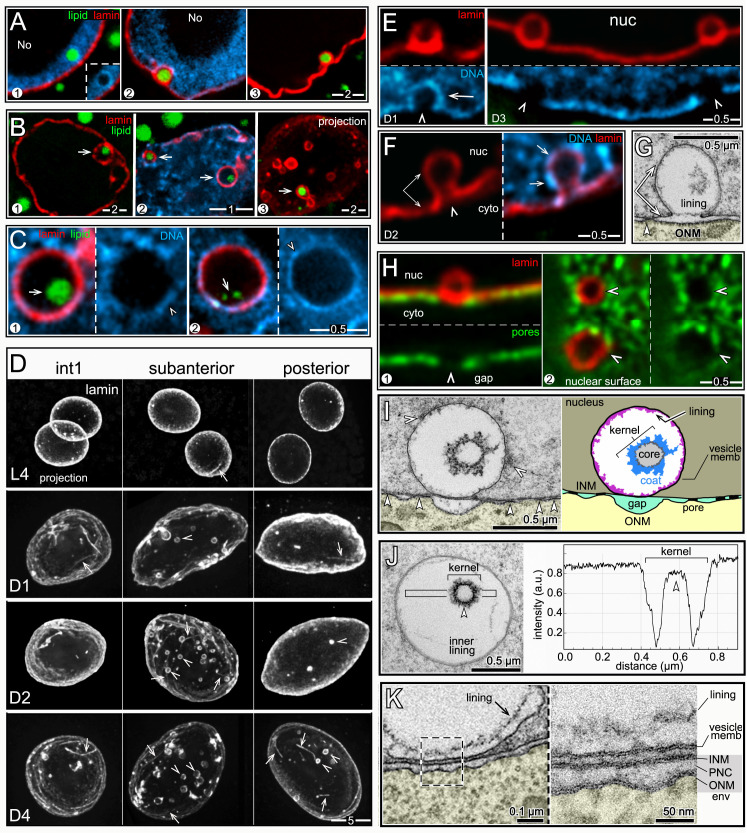
Fig 6. nLDs and lamin sacs.
(A) Examples of nLDs with and without lamin coats (red, LMN-1/lamin) in L4 (panel 1) and D1 (panels 2,3) intestinal nuclei. (B) Examples of nLDs that are within, but that do not fill, lamin sacs (arrows). The lamin sacs are closely associated with the nuclear envelope; panels 2 and 3 are tangential planes through the surfaces of D2 and D3 nuclei, respectively. Panel 3 shows a 5 μm maximum intensity z projection to illustrate the number of lamin sacs; note that only one lamin sac appears to contain lipid (arrow). (C) High magnification views of nLDs (arrows) within lamin sacs. The small, internal lipid droplets (arrows) do not appear to be directly surrounded by heterochromatin, although the lamin sac itself has a variable coating of heterochromatin (arrowheads). (D) Intestinal nuclei stained for lamin (white, LMN-1) from different regions of the intestine and at different stages as labeled; the images are maximum intensity projections showing the entire nucleus. Lamin lines (arrows) in the projection are in the nuclear interior, but nearly all of the lamin sacs (arrowheads) are just inside the nuclear envelope. (E) Images of lamin sacs in D1 and D3 nuclei. Lamin sacs in D1 nuclei have distinct coatings of heterochromatin that appear to result from inpocketings of the peripheral heterochromatin. Lamin sacs in D3 and older nuclei typically have relatively little, if any, coating of heterochromatin. (F) D2 nucleus showing a candidate precursor of a lamin sac. The image appears to show an inpocketing of the lamina (double arrow) and peripheral heterochromatin (arrows). (G) TEM of a D2 nucleus showing a candidate precursor of a lamin sac/kernel vesicle. The INM (double arrow) appears to have separated from the ONM to form an inpocketing, and there are no nuclear pores (arrowhead) at the base of the inpocketing. The membrane inpocketing has an irregular inner lining, and clumps of material appear at the outer, nucleoplasmic surface of the inpocketing. (H) D1 intestinal nuclei showing gaps (arrowheads) in the distribution of nuclear pores (green, NPP-9/RanBP2) that are coincident with the positions of lamin sacs. Panel 1 is an optical cross-section of a nucleus, and panel 2 is a tangential plane through the surface of a nucleus. (I) TEM and diagram of a kernel vesicle, or presumptive lamin sac, at a gap between pores. The kernel vesicle appears to surround a small nLD, similar to immunostained images (Fig 6C). The membrane surface of a kernel vesicle can be coated with variable clumps of material (white arrowheads in left panel), and nearly always has an irregular, inner lining of material. (J) Intensity scan through a kernel vesicle, showing the increased electron density of the lipid-like core. (K) TEM comparing a kernel vesicle membrane with the adjacent nuclear membranes. The high magnification inset shows that each of these membranes has the typical sandwich appearance of a lipid bilayer. This morphology is consistent with the proposed origin of the vesicle membrane as an inpocketing of the INM (see Fig 6G). Scale bars as indicated in microns.

