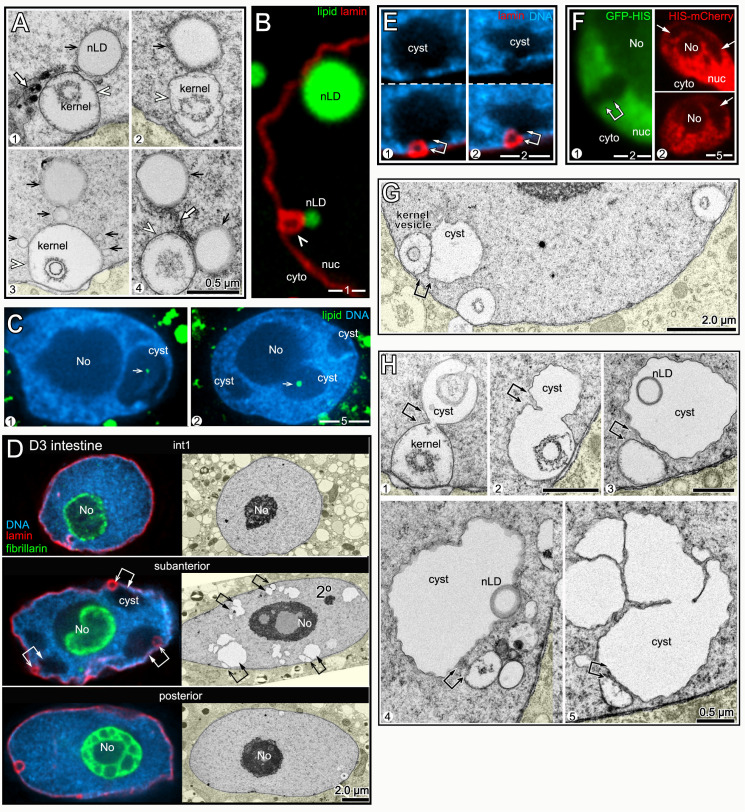
Fig 7. nLDs and nucleoplasmic cysts.
(A) TEM images of nLDs (black arrows) at the perimeter of kernel vesicles (arrowheads). Note that there are five nLDs around the kernel vesicle in panel 3. Note also nuclear tubules in panels 1 and 4 (white arrows). (B) D2 nucleus with two nLDs, one at the perimeter of a lamin sac (arrowhead) as in Fig 7A. (C) Examples of D3 nuclei with small nLDs (arrows) inside large nucleoplasmic cavities or cysts. (D) Immunostained (left column) and TEM images (right column) of D3 nuclei from the indicated regions of the intestine. Large nucleoplasmic cysts are visible in the subanterior nuclei, but not in the flanking nuclei (see also S5 and S6 Figs). By TEM, the cysts do not resemble secondary nucleoli (labeled in the subanterior nucleus), and do not stain for nucleolar markers (green, anti-fibrillarin). Double arrows in the subanterior nuclei indicate apparent pairings of cysts with lamin sacs/nuclear vesicles. (E) Examples of apparent pairings (double arrows) between cysts and lamin sacs in D3 nuclei. Note that lamin sacs, but not cysts, are adjacent to gaps in the peripheral heterochromatin. (F) Intestinal nuclei in live, D3 worms expressing one of two transgenic, fluorescent reporters for histones. The left panel shows GFP::HIS-2B (green; strain JM149), and the right panel shows HIS-24::mCherry (red; strain RW10062). Both reporters show histone-deficient regions in the nucleoplasm (arrows) consistent with the variable shapes and sizes of cysts or sac/cyst pairs. (G) TEM of D3 nucleus with three kernel vesicles, one of which is paired with a cyst. (H) TEM of D2 (panels 1,2) and D3 (panels 3–5) nuclei showing kernel vesicles paired with cysts. Panels 1 and 2 show kernel vesicle membranes continuous with cyst membranes. The cysts in panels 3 and 4 contain lipid droplets (compare with Fig 7C). The cyst in panel 5 has a cauliflower shape and appears to be fragmented.

