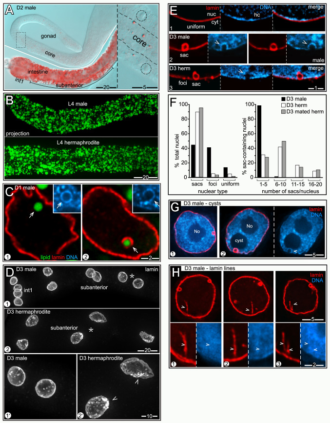
Fig 9. Male nuclei have few nLDs and appear healthier than hermaphrodite nuclei.
(A) Fat (red, oil red O) in a D2 male intestine and gonad; the inset shows the boxed region of the gonad at higher magnification with germ nuclei outlined. (B) Comparison of fat (green, BODIPY) in L4 male and L4 hermaphrodite intestines. Images are 8 μm maximum intensity z-projections. (C) nLDs (arrows) in D1 male nuclei; both have heterochromatin coats (insets), but only one has a lamin coat. (D) Nuclei in D3 male and hermaphrodite intestines stained for lamin (white, LMN-1). Panels 1’ and 2’ show higher magnifications of the subanterior nuclei indicated by asterisks in panels 1 and 2, respectively. All images are maximum intensity z-projections representing entire nuclei. Note that the male nuclei are relatively small and round and have fewer lamin sacs. Arrowheads indicate clusters of lamin sacs in the hermaphrodite nuclei. (E) Types of nuclear lamina in D3 intestinal cells, stained for lamin (red, LMN-1). The lamina can appear uniform (panel 1), contain lamin sacs (panel 2), or have foci with or without sacs (panel 3); quantified in Fig 9F. Lamin sacs in D3 male nuclei nearly always have distinct heterochromatin coats (arrowheads in panel 2), similar to younger hermaphrodite nuclei at D1 and D2. However, lamin sacs in D3 hermaphrodite nuclei usually lack distinct heterochromatin coats (arrowhead in panel 3; see also Fig 6E). (F) Quantification of nuclear types in D3 males and hermaphrodites. The numbers of nuclei scored were as follows: D3 males (784), D3 hermaphrodites (146), mated D3 hermaphrodites (215). (G) Nucleoplasmic cysts in D3 male nuclei. Male nuclei typically lack cysts (panel 1), or have only one cyst (panel 2). When present, the cysts are always associated with lamin sacs (panel 2). The arrowhead indicates the heterochromatin coat around the sac. (H) D3 male nuclei with lamin lines (arrowheads). The high magnification insets at bottom show that the lines have little apparent association with heterochromatin, by contrast with lamin lines in D3 and D4 hermaphrodites. Percentages of nuclei with lamin lines were quantified by mixing fixed D3 male and D3 hermaphrodite intestines prior to permeabilization and immunostaining; 37.4% of the male nuclei (n = 374) had at least one lamin line compared to 70.7% (n = 215) of hermaphrodite nuclei. At younger stages, the following male nuclei had lamin lines: L4 (1.5%, n = 99), D1 (18.8%, n = 80).

