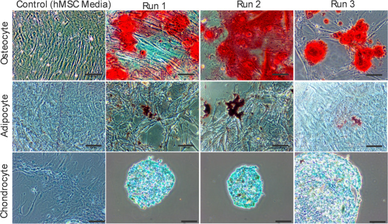
Fig 9. Stem cell induction.
Cells harvested on day seven from the bioreactor and cultured out in respective differentiation media following specialized protocols. The top row shows osteocyte, the middle row shows adipocyte, and the bottom row shows chondrocyte inductions. After specified time points based on the individual ATCC protocols cells were fixed, stained, and imaged using light microscopy. Scale bars are 100μm. The first column shows stained negative controls of respective cultures.

