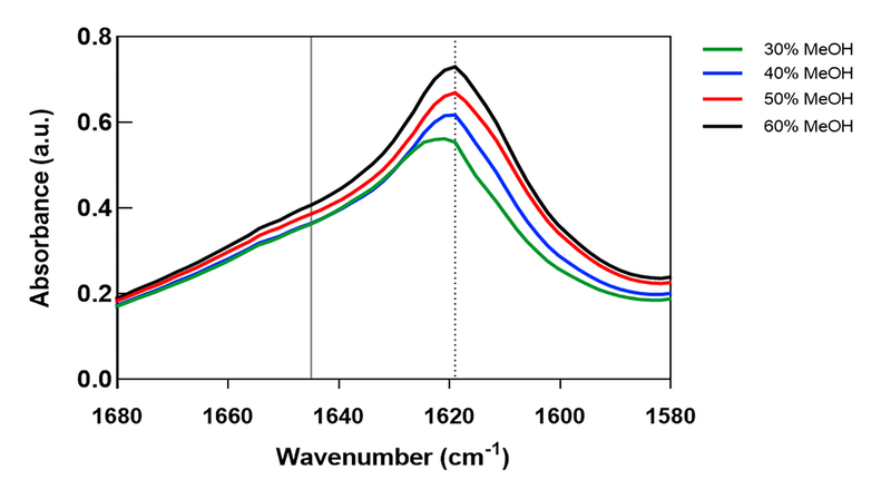
Figure 4.
Secondary structures of MeOH-annealed silk fibroin films analyzed with FTIR-ATR. β-sheet (1610–1625 cm−1) and random-coil (1640–1650 cm−1) structures are indicated by dashed and solid vertical lines, respectively. FTIR spectral data for 30%, 40%, 50%, and 60% MeOH-annealed silk films. Peak heights corresponding to β-sheet structures increase with greater MeOH-annealing concentrations.

