
Fig. 4. Modular adaptation of the OptoAAV system to different cell types.
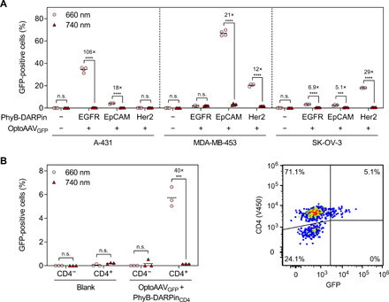
(A) Transduction of different cell lines with PhyB-DARPin adapter proteins specific for the cell surface receptors EGFR, EpCAM, and Her2. The cell lines were incubated as indicated with 50 nM PhyB-DARPin and OptoAAVGFP (MOI: 4.1 × 10⁴) in PBS supplemented with 10% FCS for 2 hours under 740- or pulsed 660-nm illumination. Afterward, cells were washed and incubated in medium under continued illumination for 46 hours before analysis of transduced (GFP-positive) cells by flow cytometry. n > 1200 cells per sample. (B) Light-controlled transduction of primary human CD4+ T cells. T cells were incubated with 50 nM PhyB-DARPinCD4 and OptoAAVGFP (MOI: 4.7 × 10⁴) in PBS supplemented with 10% FCS for 6 hours under 740- or pulsed 660-nm illumination. Afterward, cells were incubated in medium under continued illumination for 42 hours. Following staining of the cells with a V450-labeled anti-CD4 antibody, the number of transduced (GFP-positive) CD4-positive and CD4-negative cells was analyzed by flow cytometry. n > 900 cells per sample. Mean is indicated. n.s., P ≥ 0.05; ***P < 0.001; ****P < 0.0001.