
Fig. 5. Spatiotemporally controlled transduction.
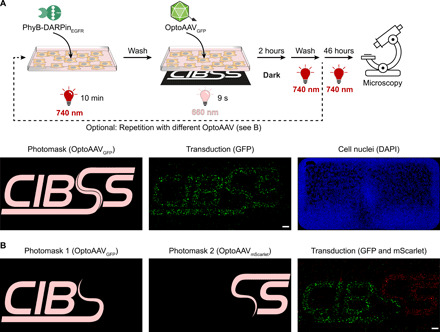
(A) Transduction with one transgene. A-431 cells were incubated with 50 nM PhyB-DARPinEGFR in PBS supplemented with 10% FCS under 740-nm light for 10 min. After washing, OptoAAVsGFP (MOI: 1.7 × 10⁴) in PBS supplemented with 10% FCS were added, and cells were illuminated for 9 s from the bottom with 660-nm light (15 μmol m−2 s−1) using the photomask with simultaneous global 740-nm illumination (3 μmol m−2 s−1) from the top. Following 2-hour incubation in the dark, cells were washed and incubated for 46 hours in medium under 740-nm light. Last, cells were fixed, DAPI-stained, and imaged by confocal microscopy. Representative images from n = 4 experiments. (B) Transduction with two transgenes. A-431 cells were spatially resolved transduced as in (A) using photomask 1 and OptoAAVGFP (MOI: 1.5 × 10⁴). Following incubation in the dark, the procedure was repeated using photomask 2 and OptoAAVmScarlet (MOI: 1.7 × 10⁴). After the second incubation step in the dark, cells were washed and incubated for 44 hours in medium under 740-nm light. Last, cells were fixed, DAPI-stained, and imaged. The DAPI image is shown in fig. S12C. Representative images from n = 2 experiments. Scale bars, 1 mm.