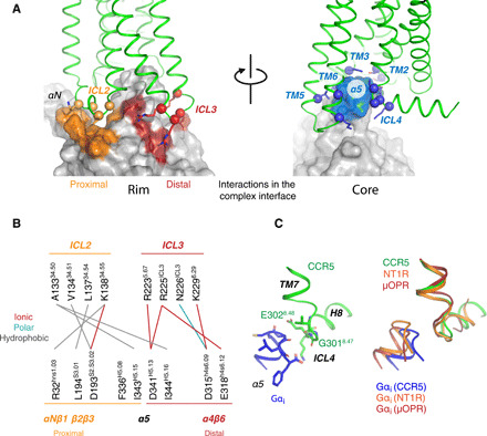
Fig. 5. Binding interfaces between CCR5 and Gαi.

(A) Binding interfaces at the rim (left) and core (right) of the complex. Each interface is colored on the surface of the G protein and interacting residues in the receptor are shown as spheres (Cα) and sticks (side chains). (B) Residue-residue interactions at the rim of the binding interface. (C) Left: Structure of the α5 hook and ICL4 in the [6P4]CCL5•CCR5 complex. Key residues are shown as sticks. Right: Comparison between the α5 hook and ICL4 in the Gi-bound receptors CCR5, neurotensin type 1 (NT1R), and μ-opioid (μOPR).
