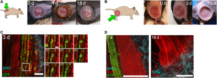
Fig. 3. PDMS-based intravital windows allow high-resolution longitudinal imaging of injury induced muscle regeneration in vivo.

(A and B) Representative photographs of a PDMS intravital imaging window maintained on the back (A) or thigh (B) muscle of a mouse for the indicated period in days (d). Brown skin coloring at day 1 in (A) is due to dermal Betadine solution. Representative of n = 9 mice implanted on the back or thigh muscles. (C) IVM of thigh muscle regeneration through a PDMS imaging window 3 days (3 d) after cardiotoxin-induced muscle injury in Pax7CreERT2;R26mTmG adult mice. All cells were labeled with membrane tdTomato fluorescent protein (Tom, red). Tamoxifen-induced Cre-mediated enhanced GFP protein expression (green) in Pax7-positive muscle stem cells and their progeny. SHG imaging shows fibrillar collagen organization in blue. Time-lapse imaging shows muscle stem cell division and migration (white asterisks) over 70 min. Scale bars, 100 μm. (D) Representative IVM images of muscle fibers 7 and 18 days after implantation in a Pax7CreERT2;R26mTmG mouse, showing resolution at the cellular scale and maintenance of excellent imaging quality over time. Scale bars, 100 μm. The small bump in the image at 7 days is due to the breathing-induced movement during image acquisition.
