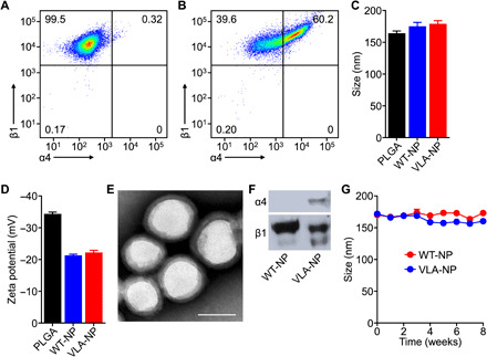
Fig. 2. Development and characterization of inflammation-targeting nanoparticles.

(A and B) Expression of integrins α4 and β1 on C1498-WT (A) and C1498-VLA (B) cells was confirmed by flow cytometry. (C and D) The average diameter (C) and surface zeta potential (D) of PLGA cores, WT-NP, and VLA-NP were confirmed by dynamic light scattering (n = 3, mean + SD). (E) Representative transmission electron microscopy image of VLA-NP (scale bar, 100 nm). (F) Western blots for integrins α4 and β1 on WT-NP and VLA-NP. (G) Size of WT-NP and VLA-NP when stored in solution over a period of 8 weeks (n = 3, mean ± SD).
