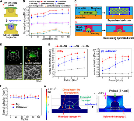
Fig. 3. Adhesive mechanism and performance of the DIAs embedded with biofluid-capturing hydrogels (H-s-DIAs) in dry and wet conditions.

(A) Schematic illustration of the s-DIA embedded with hydrogel for instantaneous capture of liquids. (B) Measurements of normal adhesion for H-s-DIA in dry (red) and underwater (blue) conditions, s-DIA in dry (violet) and underwater (sky blue) conditions, and bare hydrogel in dry (orange) and underwater (green) conditions with respect to contact time. (C) Schematic illustration of the interfacial fluid transport on the hydrogel-substrate interface for the (i) bare hydrogel before and after liquid absorption and the (ii) H-s-DIA before and after hydrogel swelling within its inner chamber for the optimized state. (D) Confocal fluorescence microscopy images of the embedded hydrogel (i) before and (ii) after the hydrogel is swelled for 2 hours. (E) Adhesive performances of the H-s-DIA adhesives in different states with varying preloads applied in (i) dry and (ii) underwater conditions. Here, the different states of the H-s-DIAs consist of H-s-DIA before contact (orange), H-s-DIA after contact for 2 hours (red), H-s-DIA (blue), and flat samples (gray). (F) Cyclic measurements of normal adhesion for H-s-DIA after 2 hours of contact time in dry and underwater conditions. (G) FEM simulation analysis of the stress distribution and structural deformation of an H-s-DIA during the application of a 2 N/cm2 preload. After the application of a weak preload, the volume of the suction chamber is effortlessly minimized by swelled hydrogel, inducing a vacuum-like state within the chamber.
