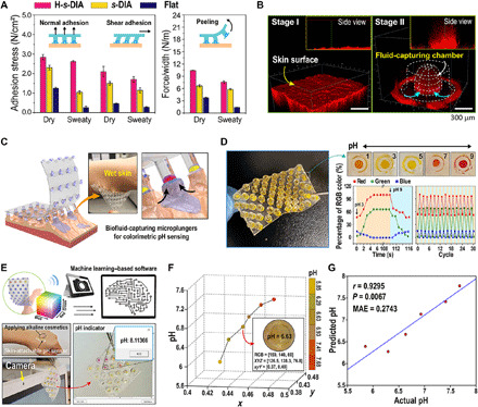
Fig. 4. Applications of the H-s-DIA patch on human skin for pH diagnosis and machine learning–based quantification.

(A) Profiles of normal, shear, and peeling adhesion of H-s-DIA against dry and sweaty pigskin. (B) Confocal fluorescence microscopy of liquid capture into the embedded hydrogel chamber of H-s-DIA [red, mixture of fluorescent dye (rhodamine 6G), water, and silicon oil]. (C) Schematic illustration of the adhesive patch with skin-attachable, biofluid-capturing suction chambers. The inset shows a representative photographic image of H-s-DIAs of 3-mm tip diameters attached to wet human skin. Photo credit: S. Baik, Sungkyunkwan University. (D) Photographic image of the diving beetle–inspired adhesive embedded with colorimetric hydrogel for reversible skincare monitoring. Time-dependent profiles of red, green, and blue color percentages and cyclic measurements of red, green, and blue color percentages for different pH values of 3 and 9 on skin. (E) Software program for quantifying pH values via supervised machine learning. The software automatically provides pH value by converting RGB values of the captured hydrogel image into x and y values. Photo credit: S. Baik, Sungkyunkwan University. (F) Scatter plot of colors (i.e., x and y values) and pH values. An example image is shown with its color and pH values. (G) Correlation between actual and predicted pH values.
