
Figure 4.
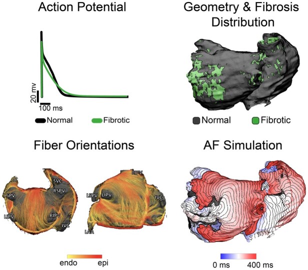
Organ-level atrial models integrate information from cell-, tissue-, and organ-level scales. For cell-level atrial electrophysiology, an example of atrial action potentials in fibrotic (green) and normal cells (black) is shown. At the tissue scale, atrial fibre architecture creates a preferential direction for wave propagation along the atrial fibres, as shown by the diffusion-tensor MRI fibre orientations acquired in explanted human atria.134 At the organ scale, personalized atrial models with patient-specific geometry and fibrotic remodelling are reconstructed from patients’ LGE-MRI scans and used in simulations of AF inducibility. Shown is an activation map of an AF episode. Modified with permission from Aronis et al.135