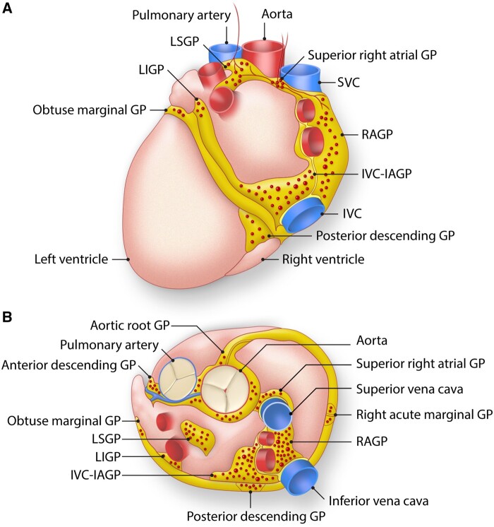
Figure 2.
ICNS. The ICNS is composed of ganglionated plexi that cluster at the hilum of the heart, close to where the PVs enter the left atrium. Anatomy of the ganglionated plexuses (GPs) that comprise the intrinsic cardiac nervous system. These GPs are typically found on the posterior (A) and superior (B) epicardial surfaces of the heart. IVC indicates inferior vena cava; IVC-IAGP, inferior vena cava-inferior atrial ganglionated plexus; LIGP, left inferior ganglionated plexus; LSGP, left superior ganglionated plexus; RAGP, right atrial ganglionated plexus; SVC, superior vena cava. Adapted from Rajendran et al., 2017.

