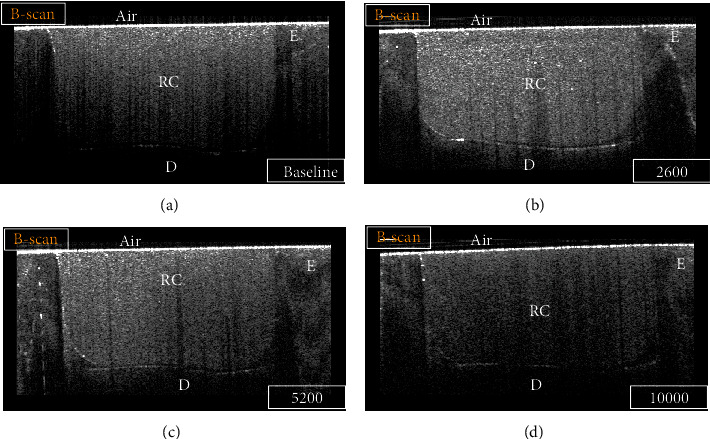
Figure 3.

B-scan for a representative specimen of the SN group at different time intervals: (a) baseline, (b) 2600 TC, (c) 5200 TC, and (d) 10000 TC. Strong backscattered reflection at the cavity floor in some regions was demonstrated on the B-scan as bright bands of pixels that were considered an interfacial gap. In contrast, regions that did not show an increase in signal intensity at the tooth-resin interface indicated no loss of interfacial seal. E: enamel; D: dentin; RC: resin composite.
