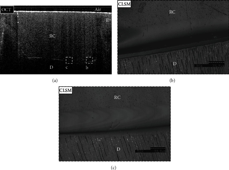
Figure 5.

Representative images for the SN group (a–c). (a) B-scan showed low backscattered reflection at the cavity floor in some areas (dotted box (b)), while other areas showed intense backscattered reflection (dotted box (c)). (b, c) Confirmatory CLSM images (×50 magnification) corresponded to the obtained OCT findings. (b) The interfacial gap was detected within the adhesive layer and corresponded to the bright band of pixels in the OCT B-scan. (c) At the same time, it showed no interfacial gap in the dentin-resin interface of other areas, which was seen as dark pixels at the cavity floor in the presented OCT B-scan. E: enamel; D: dentin; RC: resin composite.
