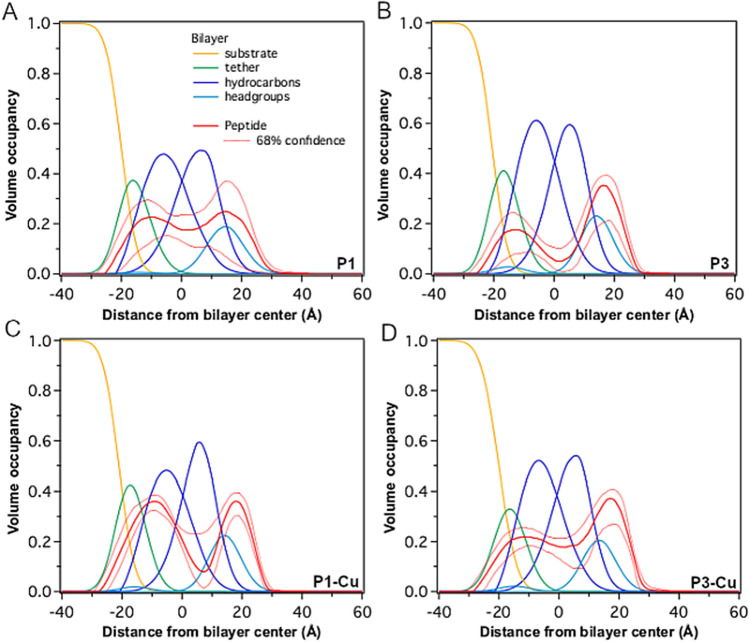
Figure 4.
Neutron reflectometry of P1 and P3 in the apo- and holo-states. The time and ensemble average spatial profiles of the various components of the tBLM (Fig. 2A) are shown as projections on the normal to the bilayer surface for (A) P1, (B) P3, (C) P1-Cu2+, and (D) P3-Cu2+. The peptides were metallated using a 1:1 stoichiometric amount of CuCl2. The inner bilayer leaflet is attached to the gold coated substrate (yellow) via molecular tethers (green). The outer leaflet of the bilayer is exposed to the aqueous compartment from which each peptide solution is injected.

