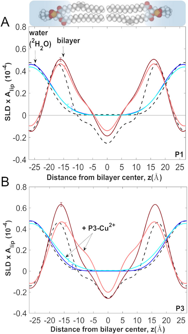
Figure 5.

Bilayer scattering length density profiles from neutron diffraction on oriented lipid multilayers. (A) The scattering length density (SLD) profiles of the bilayer are shown as projections on the bilayer normal (z-axis) for POPC (black), P1/POPC (dark red), and P1-Cu2+/POPC (pink). The peptides were metallated using a 1:1 stoichiometric amount of CuCl2. The corresponding water profiles, determined from H2O/2H2O contrast are overlaid for POPC (black), P1/POPC (dark blue), and P1-Cu2+/POPC (light blue). Peptides distribute equally on both sides of the bilayer during their incubation with liposomes and deposition on the substrate (see Methods). (B) Same as in (A) but for P3. All oriented samples were prepared in POPC at P/L = 1:25, and measured at 23 °C and 93% relative humidity achieved by using the vapor phase of saturated salt solutions. The small error bars on the curves represent the uncertainty in the profiles, which were calculated using a 95% confidence interval in the Monte-Carlo sampling of the structure factors87. Structure factors and standard deviations are given in Table S2. All profiles were determined on a per-lipid scale using structure factors calibrated to reflect the composition of the unit cell and without explicitly determining the area per lipid87.
