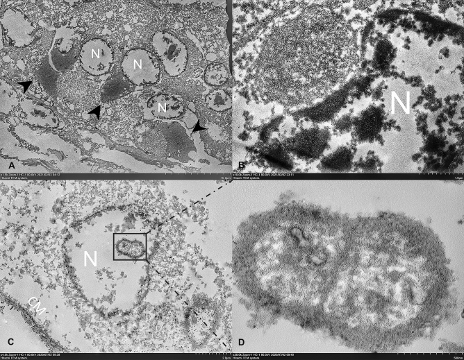
Figure 4.
Transmission electron microscopic images of reptilian ferlavirus in the snake epididymis. Representative TEM images from a big-eyed pit viper snake from colony A. (A). Intracytoplasmic, large, electron-dense viral factories (arrowheads) were observed in multiple degenerated epithelial cells represented by cellular vacuolation and a disrupted nuclear membrane (N). (B) A cytoplasmic inclusion body contains numerous pleomorphic, electron-dense viral nucleocapsid particles displacing the nucleus (N). (C) Ferlaviral ribonucleocapsid particle was seen in the nucleus (N). (D) Pleomorphic ribonuleocapsid with herringbone-like structure. Bars indicate as described in figures. CM cellular membrane.

