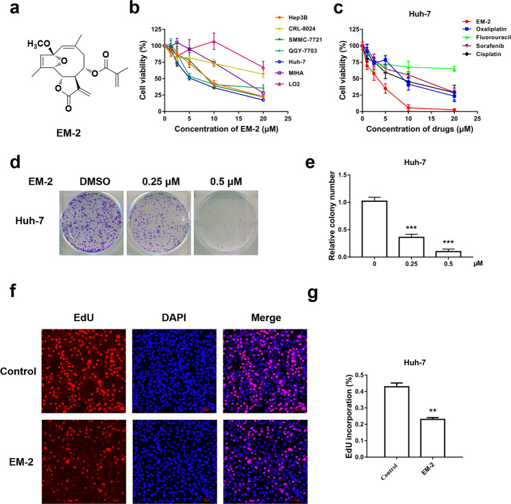
Fig. 1. Cytotoxic effect of EM-2 on human hepatocellular carcinoma cell lines.
a The chemical structure of EM-2. b Cell viability analysis of various human hepatoma cell lines and normal liver epithelial cell lines cultured in the presence of EM-2 as determined by the MTT assay. c Cell viability analysis of Huh-7 cells treated with EM-2 and various liver cancer chemotherapy drugs as determined by the MTT assay. d Huh-7 cells were seeded in 6-well plates at a density of 500 cells per well and treated with different concentrations of EM-2 (0, 0.25, and 0.5 μM) for 7 days. The results represent three independent experiments. e Statistical analysis of colony formation (*** compared with the control group, P ≤ 0.001). f The effects of EM-2 on Huh-7 cell proliferation were determined by EdU staining; the red dots represent the population of daughter cells. After exposure to EM-2 (2 μM) for 24 h followed by EdU staining, the cells were examined under a multifunctional fluorescence microscope. Scale bars, 50 μm. g Statistical analysis of the EdU staining assay (** compared with the control group, P < 0.01).

