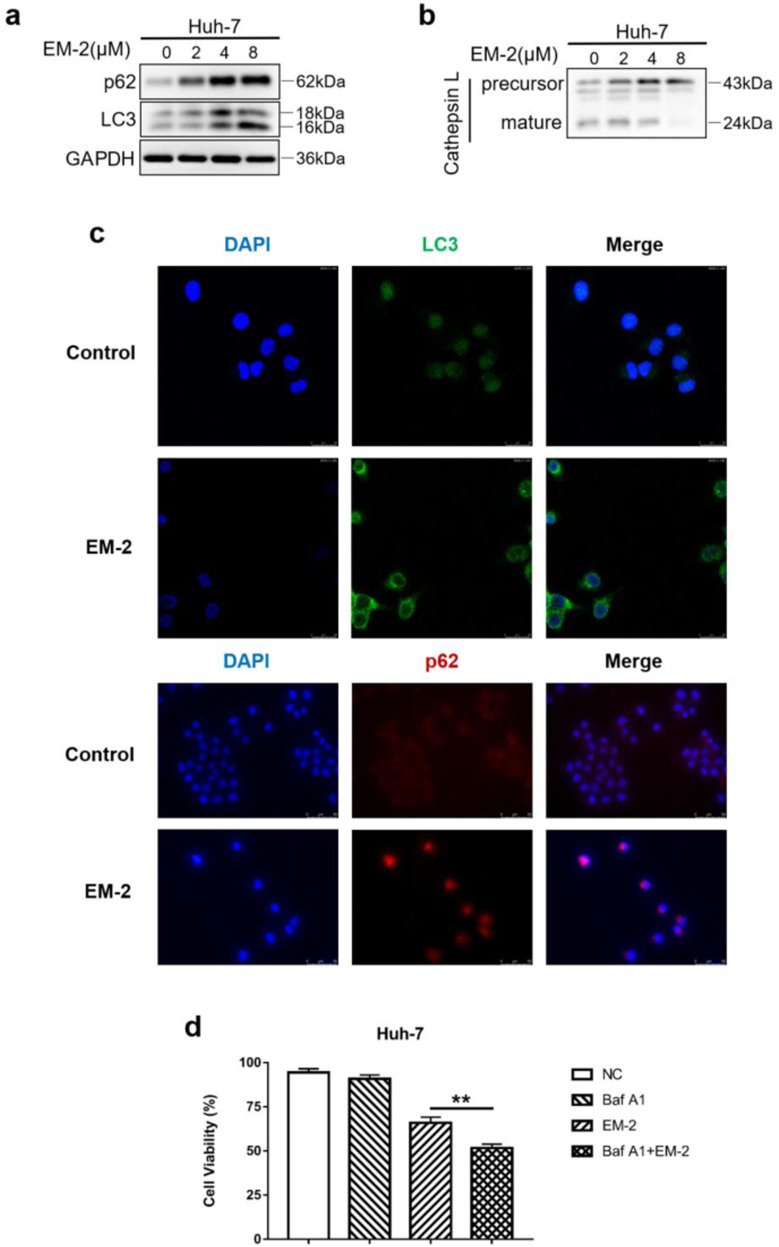
Fig. 6. EM-2 inhibited autophagy in Huh-7 cells by blocking the fusion of autophagosomes with lysosomes.
a, b Huh-7 cells were exposed to 0, 2, 4, or 8 μM EM-2 for 24 h. The protein expression levels of genes associated with autophagy were analyzed by Western blotting. c Immunostaining of LC3 and p62 in Huh-7 cells treated with different concentrations of EM-2 (0 and 8 μM). LC3 staining was imaged under a confocal laser scanning microscope, and p62 staining was imaged by an inverted fluorescence microscope. d Huh-7 cells with or without 25 μM bafilomycin A1 pretreatment were exposed to 4 μM EM-2 for 48 h, and cell viability was detected by the MTT assay. All experiments were performed in three independent replicates (*P < 0.05).

