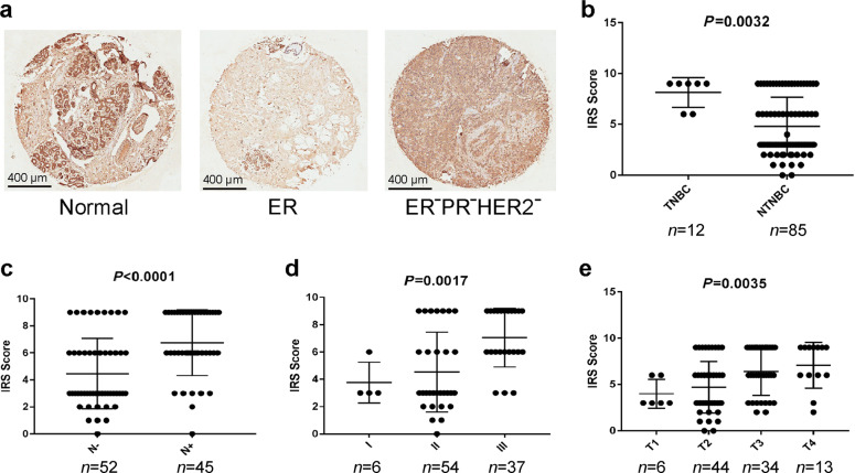
Fig. 2. FOXC1 protein was highly expressed in TNBCs and associated with breast cancer progression.
a Representative images showing the FOXC1 protein in normal tissue, ER+ breast cancer, and TNBC. Scale bar = 400 μm. b Expression of the FOXC1 protein is higher in TNBCs (n = 12) than in non-TNBCs (n = 85). c Expression of the FOXC1 protein is higher in breast cancer with positive lymph nodes (n = 45) than in breast cancer with negative lymph nodes (n = 52). d, e Expression of the FOXC1 protein was increased as breast cancer progressed. I: stage I (n = 6); II: stage II (n = 54); III: stage III (n = 37). T1: tumor T1 (n = 6); T2: tumor T2 (n = 44); T3: tumor T3 (n = 34); T4: tumor T4 (n = 13).

