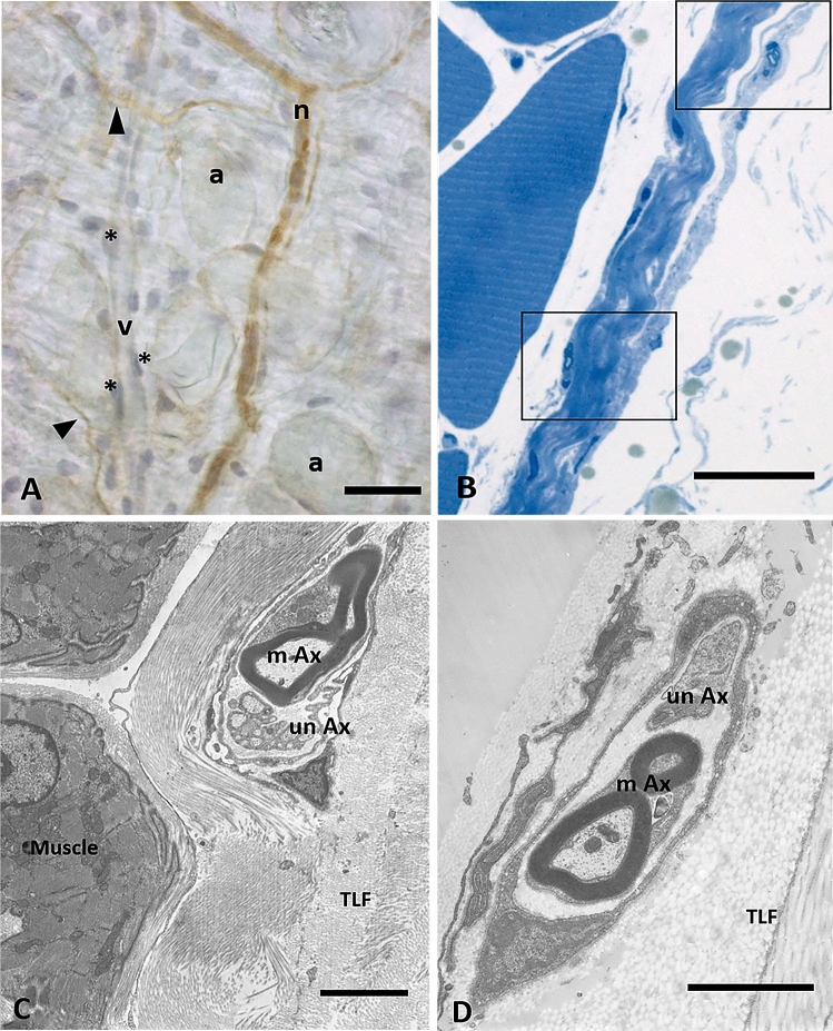
Figure 7.
Analysis of nerves inside the fascial tissue: (A) Floating thoracolumbar fascia stained with anti-S100 antibody and ematoxylin: the nervous structures are S100 positive (n: small nerve, arrows indicate single nerve fibers), whereas blood vessels are not stained (v: vessel; *: endothelial cells; a: adipocytes). (B) Semithin section of thoracolumbar fascia, whose boxes show nerve structures in the midst of collagen bundles of the fascial layers. (C) and (D): TEM images of a small nerve fiber in the inner layer (C) or in the outer layer (D) of the TLF, with both myelinc and unmyelinic axons. m: muscle; TLF: thoracolumbar fascia; mAx: myelinic axon; unAx: unmyelinic axon. Scale bars: (A) and (B) 30 µm; (C) 3 µm; (D) 2 µm.

