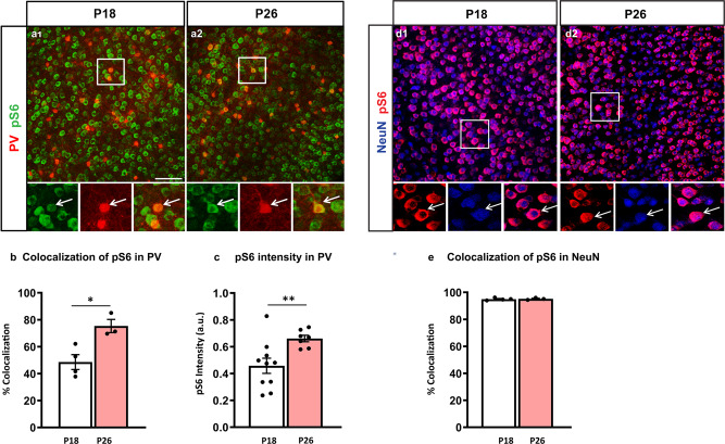
Fig. 1. pS6 expression levels increase specifically in PV cells between the second and fourth postnatal weeks.
a Coronal sections of mouse somatosensory cortex immunostained for pS6 (green) and PV (red) at P18 (a1) and P26 (a2). b Number of PV cells expressing detectable levels of pS6 increases during the second to fourth postnatal week (Welch’s t-test, *p = 0.0152). Number of mice; P18, n = 4; P26, n = 3. c Mean pS6 intensity in individual PV cells is significantly higher at P26 than at P18 (Welch’s t-test, **p = 0.0061). Number of mice; P18, n = 10; P26 n = 7. d Coronal sections of mouse somatosensory cortex immunostained for pS6 (red) and NeuN (blue) at P18 (d1) and P26 (d2). e Percentage of colocalization of pS6 and NeuN is not significantly different between the two developmental ages (Welch’s t-test, p = 0.7663). Number of mice; P18, n = 4; P26, n = 3. Scale bars in a1-a2, d1-d2, 75 µm. Data represent mean ± SEM. Source data are provided as a Source Data file.

