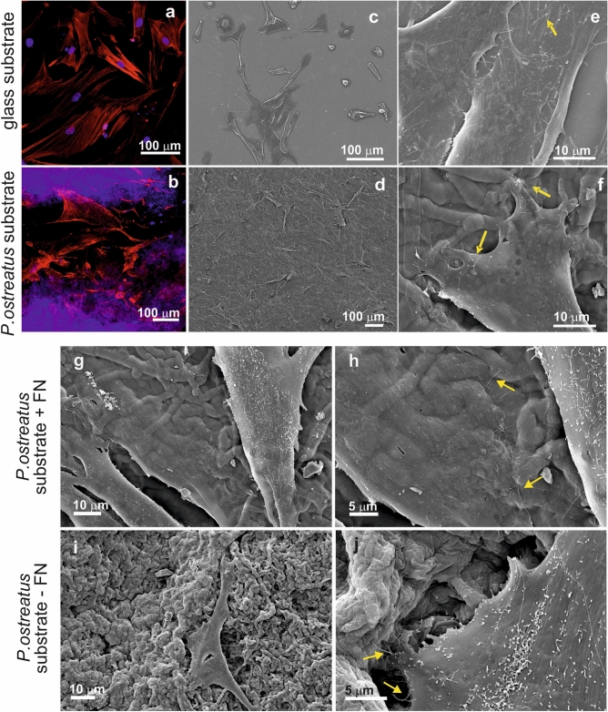
Figure 5.
Confocal (a) and SEM micrographs (c, e) of HDFa cells grown for 48 h onto a FN-coated glass coverslips. Confocal (b) and SEM micrographs (d, f) of HDFa cells grown for 48 h onto a FN-coated P. ostreatus substrate. Nuclei are stained with DAPI (visible in blue), while the actin fibers are stained with Alexa Fluor Phalloidin 488 (highlighted in green). Filopodia extending from the cytoplasm of the attached cells are indicated with yellow arrows. (g–j) Effect of the scaffold coating onto primary cell growth. HDFa cells are seeded onto P. ostreatus scaffolds, either coated with fibronectin (g, h) or uncoated (i, j). Filopodia extending out of the attached cells are highlighted with yellow arrows.

