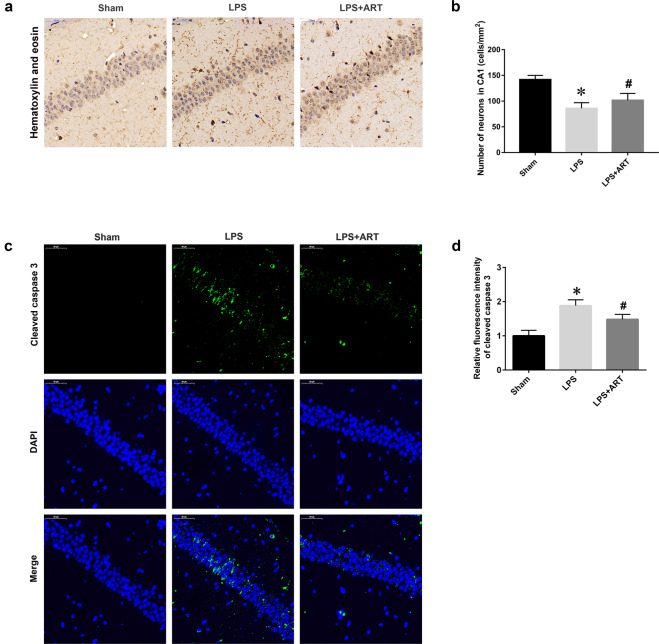
Fig. 2. Artemisinin ameliorated neuronal cell death in an LPS-induced murine sepsis model.
a Immunohistochemical staining was used to examine neuronal survival in the CA1 area of the hippocampus. Magnification: ×200. b Quantitative analysis of the data in a. c Representative images of cleaved caspase 3 staining. Magnification: ×200. d Statistical analysis of the relative fluorescence intensity of cleaved caspase 3. *P < 0.05 versus sham group mice. #P < 0.05 versus LPS-treated mice. n = 4–6.

