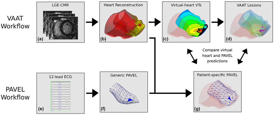
FIGURE 1.

Workflows of VAAT and PAVEL, and comparative analysis. VAAT Workflow: (A) From LGE-MRI stack for a patient, the myocardium is segmented into scar and grey zone tissues. (B) Personalized 3D virtual hearts are reconstructed from the segmented data and electrophysiological information is incorporated. (C) An endocardial activation map of the infarct-related VTs in a virtual heart. (D) VAAT-predicted ablation targets are marked by purple circles on the left ventricular endocardial surface. PAVEL Workflow: (E) The eight-lead ECGs of an induced monomorphic VT were recorded, and the other four leads (Lead III, aVF, aVL, aVR) were computed. The user can edit the onset of the 120 ms window (rectangle box) if correction is necessary. (F) A blue triangle that indicates the estimated VT exit location on a generic LV endocardial surface using PAVEL. (G) The PAVEL-predicted VT exit site on the generic LV endocardial surface was projected onto a patient-specific LV endocardial surface obtained from the personalized 3D digital heart. Comparative Analysis: we do not expect VAAT predicted ablation targets and PAVEL-predicted exit sites to necessarily co-localize; details in the text
