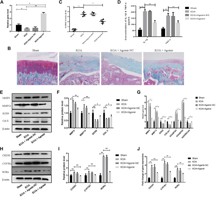
FIGURE 6.
MiR-10a-3p improves cartilage degeneration in KOA model rats. (A) Detection of transfection efficiency of miR-10-3p agomir by qRT-PCR. (B) Saffron Red & Fast green staining of cartilage tissue, 100×, Scale bar = 100 μm. (C) OARSI scores of cartilage tissue in different groups (n = 5). (D) Concentration of IL-1β and TNF-α in serum were detected by ELISA. (E,F) MMP3, MMP13, SOX9, and Collagen II expression in cartilage tissue were analyzed via western blotting. The band intensity was quantified by normalizing to β-actin (n = 3). (G) MMP3, MMP13, SOX9, COL2α1, ADAMTS4, ADAMTS5, and Aggrecan expression in cartilage tissue were analyzed via qRT-PCR. Quantitative data were presented as mean ± SD. (H,I) CH25H, CYP7B1, and RORα expression in cartilage tissue were analyzed via western blotting. The band intensity was quantified by normalizing to β-actin (n = 3). (J) CH25H, CYP7B1, and RORα expression in cartilage tissue were analyzed via qRT-PCR. Quantitative data were presented as mean ± SD. *p < 0.05, **p < 0.01.

