Fig. 1.

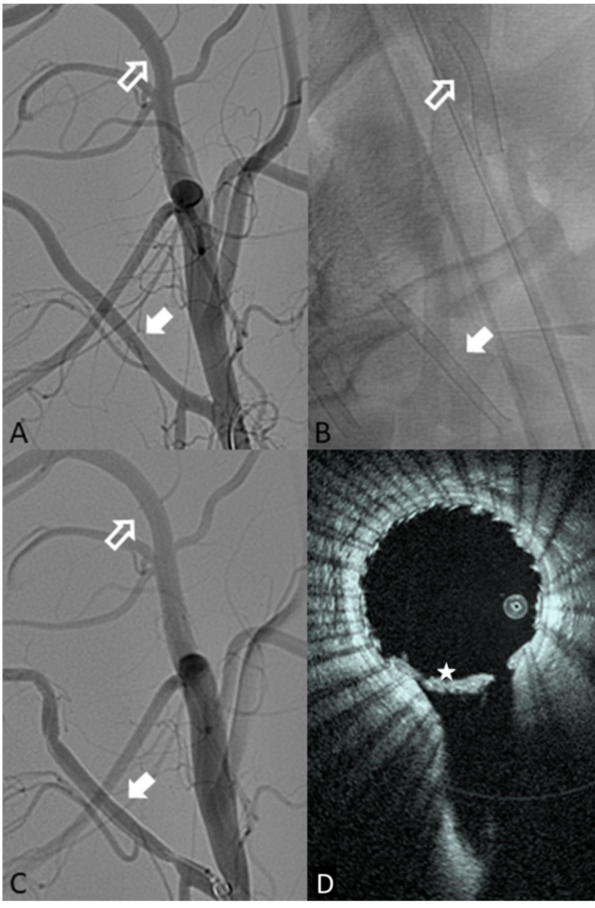
A Pre-implant DSA showing the internal maxillary target location (hollow white arrow) and the ascending pharyngeal artery target zone (solid white arrow). B Single-shot fluoroscopic image of both devices once deployed (IMAX—hollow arrow; APA—solid arrow). C Post-implant example DSA of both locations deployed (IMAX—hollow arrow; APA—solid arrow). D HF-OCT slice from the APA side branch, the star (*) shows a large amount of thrombus seen on HF-OCT but not DSA
