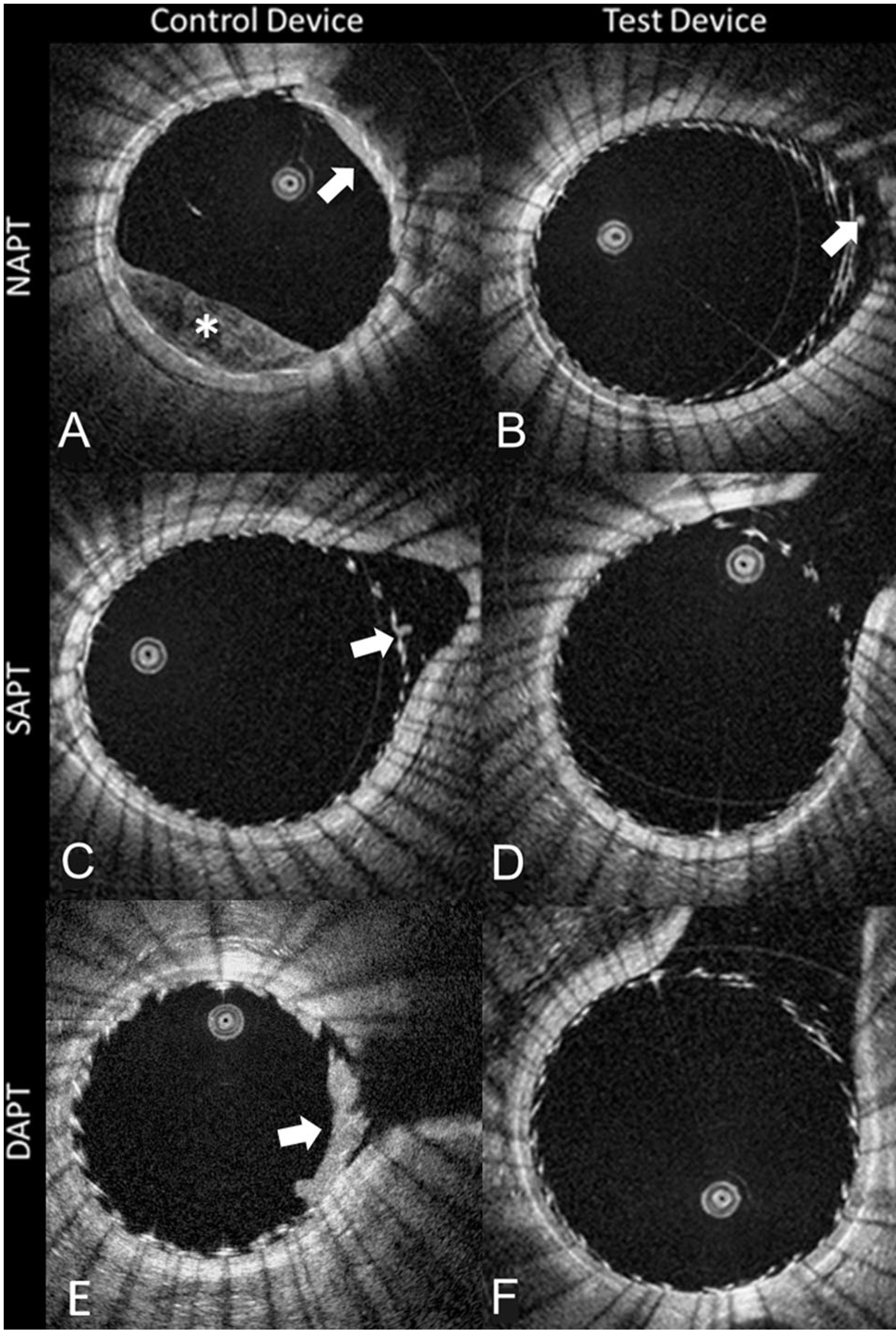
Fig. 2.

A single OCT slice derived from each of the 6 testing conditions. The first column (A, C, E) shows the control device (p-48MW), while the second column (B, D, F) shows the test device (p48MW-HPC). Along the rows shows the different anti-platelet therapy groups. Of note, only conditions A, B, C, and E show an appreciable burden of clot (white arrows), confirming that the combination of the test device and aspirin to remove almost all the clot. The (*) in panel A shows incomplete blood clearance in the lumen (NAPT; no anti-platelet therapy, SAPT: single anti-platelet therapy, DAPT; dual anti-platelet therapy)
