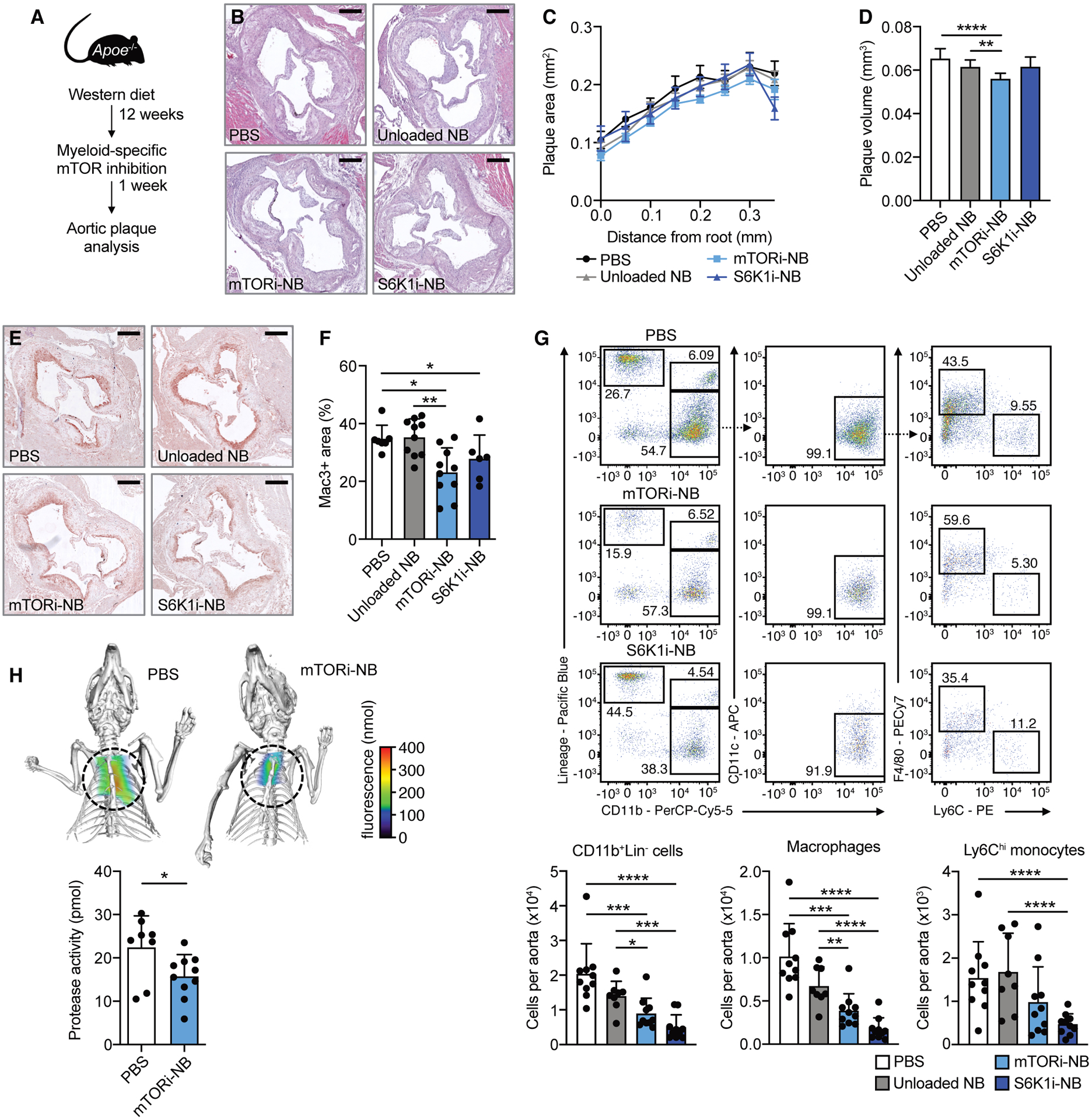
Fig. 1. Myeloid-specific mTOR inhibition reduces atherosclerotic plaque inflammation.

Apoe−/− mice were fed a Western diet for 12 weeks, followed by 1 week of treatment, while continuing the diet. Treatment consisted of 4 intravenous injections of PBS, mTORi-NB (rapamycin at 5 mg/kg), S6K1i-NB (PF-4708671 at 5 mg/kg) or unloaded nanobiologics (NB, at a comparable dose). See schematic in (A). (B) Representative images of H&E-stained aortic roots, scale bar = 250 μm. (C) Histologic quantification of plaque area at set distances from the aortic root, presented as mean ± SEM (n = 6–10 mice/group). (D) Lesion volume was calculated as area under the curve in C. (E) Representative Mac3-stained aortic roots (scale bar = 250 μm) and (F) quantification of Mac3+ area of treated mice (n = 6–10 mice/group). (G) Representative flow cytometry plots and quantification of CD11b+Lin− cells, macrophages (CD11b+Lin− CD11c−F4/80+Ly6Clo) and Ly6Chi monocytes (CD11b+Lin−CD11c−F4/80−Ly6Chi) in the aorta (n = 8–10 mice/group). (H) FMT/CT imaging of protease activity in the aortic root of PBS or mTORi-NB-treated mice (n = 8–10 mice/group). Experiments were performed once. Data are presented as mean ± SD unless otherwise stated. ANOVA with Dunnett’s correction was used in D, non-parametric Mann-Whitney U tests were applied in F, G and H. *P < 0.05, **P < 0.01, ***P < 0.001, ****P < 0.0001.
